YAG Laser Capsulotomy
by Dr Rupal Morjaria
Posterior capsular opacification is the formation of thin layer of scar tissue that develops behind the lens implant in a small proportion of patients after cataract surgery.
What is posterior capsular opacification (PCO)?
When you have cataract surgery, an artificial lens implant is placed inside the lens capsule. Posterior capsular opacification is the formation of thin layer of scar tissue that develops behind the lens implant in a small proportion of patients after cataract surgery. PCO can occur between 1-10 years after your cataract operation and is the most common cause of reduction in vision after successful cataract surgery.
How can PCO be treated?
PCO can be very successfully treated by a procedure called YAG laser capsulotomy. The procedure is performed very commonly and is painless. You will require drops to dilate your pupils and anaesthetic eye drops before the procedure.
YAG laser is performed to clear the cloudy film by making a small opening in the capsular bag. On average, the process takes 10 minutes. It is a safe and successful treatment and nearly never needs to be repeated. After the procedure, you will typically be given steroid drops to put them in the eye for a few days.
What are the risks of the procedure?
YAG laser capsulotomy is a very safe procedure. Immediately after the procedure you may see floaters which would gradually improve. This is a common symptom and is nothing to worry about. Commonly, there can be raised pressure in the eye, immediately after the procedure or inflammation inside the eye. Dr Morjaria will give you drops after the procedure to reduce the risk of this happening.
Less common risks are of seeing flashes or persistent floaters. Serious complication such as retinal damage or movement of your intraocular lens are rare.
How quickly will I recover?
People may notice a difference immediately or as soon as the dilating drops wear away. You can carry on with most routine activities immediately after your laser.
What tests are needed?
OCT Scan - Optical Coherence Tomography - This looks for damage to the photoreceptors at the back of the eye. Visual field testing - This looks for a reduction in visual fields which is common in retinitis pigmentosa. Electrodiagnostics - This is a test where wires are attached around the eye and the electrical response after your eyes are stimulated in different light conditions are measured. It is a valuable tool, to look at the function of the photoreceptors in the eye. Genotyping - A blood test can be performed to look at your genetic code for a definite diagnosis. This will help us understand the exact type or retinitis pigmentosa which will allow Dr Mojaria to advise you on the likelihood and severity of any visual loss that may occur.
Fluorescein Angiogram - You may need additional tests that involve having a yellow dye injected into your hand to take pictures of the blood circulation in your eye. This will allow Dr Morjaria to assess for areas of the eye that are not getting enough oxygen or areas that need laser treatment to prevent bleeding in your eye.

Locations
A Clinic Near You
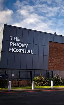
The Priory Hospital, Priory Road, Birmingham, B5 7UG
Chamberlain Clinic, 81 Harborne Road, Edgbaston, Birmingham, B15 3HG
Spire Little Aston Hospital, Little Aston Hall Drive, Sutton Coldfield, B74 3UP
6 Church Street, Oakham, Leicester, LE19 1SJ

Birmingham
Priory Hospital

Edgbaston
Chamberlain Clinic

Sutton Coldfield
Spire Little Aston Hospital

Leicester
Coe & Coe

