Diabetic Retinopathy
by Dr Rupal Morjaria
Diabetic Retinopathy is the fifth common cause of blindness in the world and a major cause of blindness.
Diabetic retinopathy is the fifth common cause of blindness in the world and a major cause of blindness among working population in developed countries. Diabetic retinopathy is treatable and vision loss can be prevented or delayed if treated early.
Diabetic complications in the eye occur due to the effects of poor blood sugar control together with other risk factors such as high blood pressure, high cholesterol & smoking. It can result in mild diabetic changes such as a few haemorrhages that do not require treatment but monitoring.
More severe disease causes bleeding or new vessels in the eye called proliferative diabetic retinopathy (PDR) or fluid build-up in the camera film of the eye (diabetic macula oedema, DMO). Both of these require urgent treatment.
Treatment
Diabetic Retinopathy used to be traditionally graded as mild (R1) moderate (R2) or severe (R3), but several different classification systems exist. If you have a mild or moderate disease, changes can be made in your diet, lifestyle and medication to reverse these changes.
Dr Morjaria will advise you on this. However, once you have severe diabetic retinopathy urgent treatment is usually required to prevent or slow down sight loss.
Dr Morjaria will tailor your treatment for diabetic retinopathy considering your general health and lifestyle. It will include injections to the back of the eye to slow down the growth of new vessels with anti-VEGF (vascular endothelial growth factor) drugs such as ranibizumab (Lucentis), aflibercept (Eylea) and bevacizumab (Avastin) together with laser to the areas of the eye that are not receiving sufficient oxygen.
Laser treatment (panretinal photocoagulation, prp) to these areas helps to divert the essential resources to areas of the retina (camera film) that are the most important for your reading vision.
Oedema
Macula oedema affects the macula region of the retina. The macula is the region of the retina used for sharp, clear vision when reading or focusing on an image. Macula oedema is the condition when you get fluid deposition in the retinal layers in the macula region.
This can result in symptoms of difficulty reading and loss of vision. Treatment of macula oedema includes gentle laser or injections to the back of the eye. The injections needed may be anti-VEGF drugs or steroid implants depending on how long you have had the condition and how much treatment you have had.
Diabetes and Glaucoma
People who suffer with diabetes are at a higher risk of developing glaucoma. This is when the pressure in the eye is too high and can cause damage to the nerve sending messages to the brain. If this is not detected early, it can cause changes in your field of vision. It is important that the doctor monitoring your eyes for diabetes also checks the pressure of the eye and the nerve at the back of the eyes.
Diabetes and Cataract
Cataract is a cloudy lens. The lens helps to focus images from the outside world onto a small part of the camera film called the fovea. As people get older their lens gets thicker and ‘yellower’. Having fluctuating blood sugars can speed up this process and people with diabetes may require cataract surgery earlier.
Dr Morjaria specialises in seeing people with diabetes and cataracts. She is thorough to take additional steps to avoid swelling and inflammation after surgery. Dr Morjaria fully assesses the eye for risks of post-operative macular oedema before her patients undergo surgery. People with diabetes may need additional treatments during or after surgery to help them get the best outcome. People with diabetes may need additional treatments during or after surgery to help them get the best outcome. Sometimes additional injections of anti-VEGF agents or steroids are needed at the time of cataract surgery to help get the best outcome. Dr Morjaria can discuss the risks and benefits of these treatments. Tests you may need:
What is a Fluorescein Angiogram (FA)?
-
A fluorescein angiogram is a test to look at the blood vessels in the back of your eye (retina).
-
A special yellow dye (fluorescein) is injected into a vein in your arm.
-
A camera with a special filter then takes rapid photographs of your retina as the dye travels through your eye’s blood vessels.
Why is it done?
To check for leakage, blockages, or abnormal blood vessels.
Helps in diagnosing conditions like:
Diabetic eye disease
Macular degeneration
Retinal vein/artery occlusions
What happens during the test?
-
Eye drops are given to dilate (widen) your pupils.
-
A small amount of dye is injected into your arm.
-
You may feel a brief warm sensation after injection.
-
A series of photos are taken over a few minutes.
-
After the test, your skin may look slightly yellow and your urine may turn bright yellow for up to 24 hours (this is normal).
What is OCT (Optical Coherence Tomography)?
OCT is a quick, non-invasive eye scan that uses light waves (not X-rays) to create high-resolution cross-section images of your retina. Think of it like an ultrasound with light.
Why is it done?
-
To measure the thickness and layers of the retina.
-
Helps in detecting and monitoring:
-
Macular degeneration
-
Diabetic macular edema
-
Glaucoma
-
Retinal swelling
What happens during the test?
-
You sit in front of the OCT machine and rest your chin on a support.
-
You look at a small target light.
-
The scanner takes images in a few seconds – no injections or touching the eye.
Key Differences
-
FA: Involves an injection and photographs the blood flow in the retina.
-
OCT: No injection, uses light to scan and map the layers of the retina.
Both tests give your doctor detailed information to help diagnose and manage eye conditions effectively
Locations
A Clinic Near You
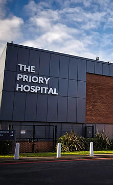
The Priory Hospital, Priory Road, Birmingham, B5 7UG
Chamberlain Clinic, 81 Harborne Road, Edgbaston, Birmingham, B15 3HG
Spire Little Aston Hospital, Little Aston Hall Drive, Sutton Coldfield, B74 3UP
6 Church Street, Oakham, Leicester, LE19 1SJ

Birmingham
Priory Hospital

Edgbaston
Chamberlain Clinic

Sutton Coldfield
Spire Little Aston Hospital

Leicester
Coe & Coe

